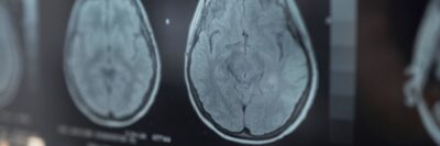

Why Rezilir Health Uses NeuroQuant MRI, qEEG, and Cognitive Testing
When a patient tells us, “My memory just isn’t what it used to be,” the answer is rarely found in a single test.
Our approach to cognitive health is based on a fundamental principle: the brain should be evaluated from more than one perspective.
A standard office cognitive screen can tell us how someone performs on a particular day. An MRI can show us what the brain looks like structurally. A qEEG can tell us something entirely different—how the brain is functioning electrically.
Each provides a different piece of information.
That is why, when clinically appropriate, we may combine NeuroQuant® volumetric MRI, quantitative EEG (qEEG) brain mapping, and objective cognitive testing such as CNS Vital Signs (CNSVS), the Montreal Cognitive Assessment (MoCA), or the Mini-Mental State Examination (MMSE).
The goal is not simply to obtain more testing. The goal is to develop a more complete picture of the brain.
Step One: NeuroQuant MRI — Looking at Brain Structure
A conventional brain MRI is an important part of evaluating cognitive changes because it allows clinicians to examine brain anatomy and identify findings such as vascular disease, prior strokes, white-matter changes, tumors, hydrocephalus, microhemorrhages, and patterns of cerebral atrophy.
Current clinical guidelines recommend structural brain imaging, preferably MRI when available and appropriate, as part of the evaluation of patients with suspected cognitive impairment.
NeuroQuant adds another dimension to that MRI.
NeuroQuant is FDA-cleared software that analyzes MRI images and automatically measures the volumes of specific brain structures. Rather than relying exclusively on visual interpretation, it provides quantitative measurements that can be compared with reference percentile data.
This can be particularly useful when examining structures involved in memory and cognition, including the hippocampus and other cortical and subcortical regions.
In other words, instead of simply asking: “Does the MRI look abnormal? We can also ask: “How much tissue is present in specific regions of this patient’s brain, and how does that compare with reference data?”
NeuroQuant also allows comparison across time when appropriate imaging is available, giving clinicians another way to evaluate whether structural changes are occurring.
Importantly, NeuroQuant does not diagnose Alzheimer’s disease by itself. Brain volume changes are not specific to a single disease, and NeuroQuant results must be interpreted alongside the radiologist’s assessment, the patient’s clinical history, examination, laboratory findings, cognitive testing, and other biomarkers when indicated.
Step Two: qEEG — Looking at How the Brain Is Functioning
While an MRI provides information about the brain’s structure, a quantitative EEG (qEEG) provides a different perspective by evaluating patterns of the brain’s electrical activity.
A standard EEG records electrical activity through electrodes placed on the scalp. qEEG takes this information a step further by using computerized analysis to evaluate brain-wave frequencies, their relative distribution, and patterns of electrical activity across different regions of the brain.
The qEEG findings can then be compared with a normative database, allowing the patient’s brain-wave patterns to be evaluated in the context of relevant characteristics such as age, sex, and handedness (right- or left-hand dominance). This helps identify areas where electrical activity may differ from expected patterns for a comparable population.
We use qEEG as one component of a comprehensive cognitive and neurological evaluation because it provides functional information that structural imaging alone cannot provide.
In simple terms:
- NeuroQuant MRI helps us evaluate brain anatomy and structural changes.
- qEEG helps us evaluate patterns of electrical activity and electrophysiologic function.
Together, these tests provide complementary information: one examines what the brain looks like, while the other helps us understand how the brain is functioning electrically.
Importantly, qEEG is an adjunctive tool. It does not independently diagnose Alzheimer’s disease, mild cognitive impairment, or another form of dementia. Its findings are interpreted alongside imaging, cognitive testing, clinical history, examination, and other relevant diagnostic information.
Step Three: Cognitive Testing — Measuring What the Patient Can Actually Do
The third piece of the evaluation brings us back to the patient. Ultimately, we need to know how the brain is performing.
Depending on the clinical situation, this may involve tools such as CNS Vital Signs, MoCA, or MMSE.
These tests approach cognition somewhat differently.
CNS Vital Signs is a computerized neurocognitive assessment that can provide information across multiple cognitive domains. In clinical practice, this can help create a cognitive performance profile rather than relying exclusively on a single global score.
The MoCA is a widely used screening instrument that assesses several cognitive domains and is particularly useful when mild cognitive impairment is a concern.
The MMSE is another established cognitive screening instrument and may be useful in clinical settings and for following patients longitudinally.
No cognitive screening test should be interpreted in isolation. Performance can be influenced by education, language, culture, hearing or vision difficulties, anxiety, depression, sleep, medications, fatigue, and other medical conditions.
When screening results do not adequately explain a patient’s symptoms—or when the clinical picture is complex—formal neuropsychological evaluation may be appropriate.
Why Combine the Three?
This is where the evaluation becomes much more informative.
Imagine three different windows into the same brain:
- NeuroQuant MRI: What does the brain look like structurally?
- qEEG: How is the brain functioning electrically?
- Cognitive testing: How is the patient performing cognitively?
Sometimes those findings line up.
For example, a patient may demonstrate memory impairment on cognitive testing, reduced hippocampal volume on quantitative MRI, and electrophysiologic abnormalities on qEEG.
Concordant findings can strengthen the rationale for additional evaluation and help us understand the patient’s cognitive profile.
But sometimes the tests do not agree—and that can be equally important.
A patient may report significant cognitive symptoms while MRI volumes remain relatively preserved. Another patient may have structural changes but perform surprisingly well on cognitive testing. Someone else may demonstrate functional abnormalities without a clear structural correlate.
Those discrepancies are not necessarily failures of testing. They are clues and tell us that we need to look deeper.
The Value of Establishing a Baseline
There is another reason we value objective testing: cognitive medicine is longitudinal medicine.
- One measurement is a snapshot.
- Repeated measurements can become a trajectory.
Establishing baseline measurements of brain structure and cognitive performance allows us to ask a much more meaningful question later:
Has something actually changed?
Rather than relying only on statements such as “I think my memory is worse” or “My family thinks I am doing better,” we can compare current findings with previous objective measurements.
That does not eliminate clinical judgment. It strengthens it.
Putting the Pieces Together
In practice, a single MRI measurement, brain-wave pattern, laboratory result, or cognitive score should not define a patient.
The evaluation of cognitive decline requires integration.
- History matters.
- Neurologic examination matters.
- Sleep matters.
- Medications matter.
- Metabolic and vascular health matter.
- Mental health matters.
- Laboratory findings matter.
- Imaging matters.
- And objective cognitive performance matters.
NeuroQuant MRI, qEEG, and cognitive testing allow us to examine different dimensions of the same extraordinarily complex organ.
Structure. Function. Performance.
When those pieces are interpreted together—and placed within the context of the individual patient—they can help us move beyond the question:
“Is there something wrong with my memory?”
And toward more useful questions:
- What pattern are we seeing?
- What could be contributing to it?
- What additional evaluation is warranted?
And how can we objectively follow this patient over time?
That is the purpose of comprehensive cognitive assessment at Rezilir Health: not simply collecting more data but connecting the data so that we can better understand the person behind it.
References
- Atri et al. — DETeCD-ADRD “Executive Summary…for primary care” — Real. The correct publication is Alzheimer’s & Dementia 2025;21(6):e14333 (PMID 39713942), not 2024. The lead author on the primary-care executive summary is Atri, as listed. [1]
- Atri et al. — DETeCD-ADRD “Validated Clinical Assessment Instruments” — Real. Published in Alzheimer’s & Dementia 2025;21(1):e14335 (PMID 39713939). Your reference dates it 2024; it appeared in the 2025 volume. [2]
- Brewer, Magda, Airriess & Smith — “Fully-Automated Quantification of Regional Brain Volumes…” — Real and accurate. AJNR Am J Neuroradiol 2009;30(3):578–580 (PMID 19112065); DOI matches. [3]
- Dickerson et al. — DETeCD-ADRD “Executive Summary…for specialty care” — Real. Alzheimer’s & Dementia 2025;21(1):e14337 (PMID 39713957); Dickerson is the correct lead author for the specialty-care summary. Again the year is 2025, not 2024. [4]
- Islam et al. — “Accuracy of the MoCA for Detecting MCI: A Systematic Review and Meta-Analysis” — Real and accurate. Alzheimer’s & Dementia, published 2023 (PMID 36934438). [5]
- Shaughnessy & Weintraub — “The Role of Neuropsychological Assessment…” — Real. Alzheimer’s & Dementia 2025 (PMID 39732508). Note the first author’s full initials are Shaughnessy LW; the article is part of the DETeCD-ADRD series. Your reference lists 2024, but it appeared in the 2025 volume